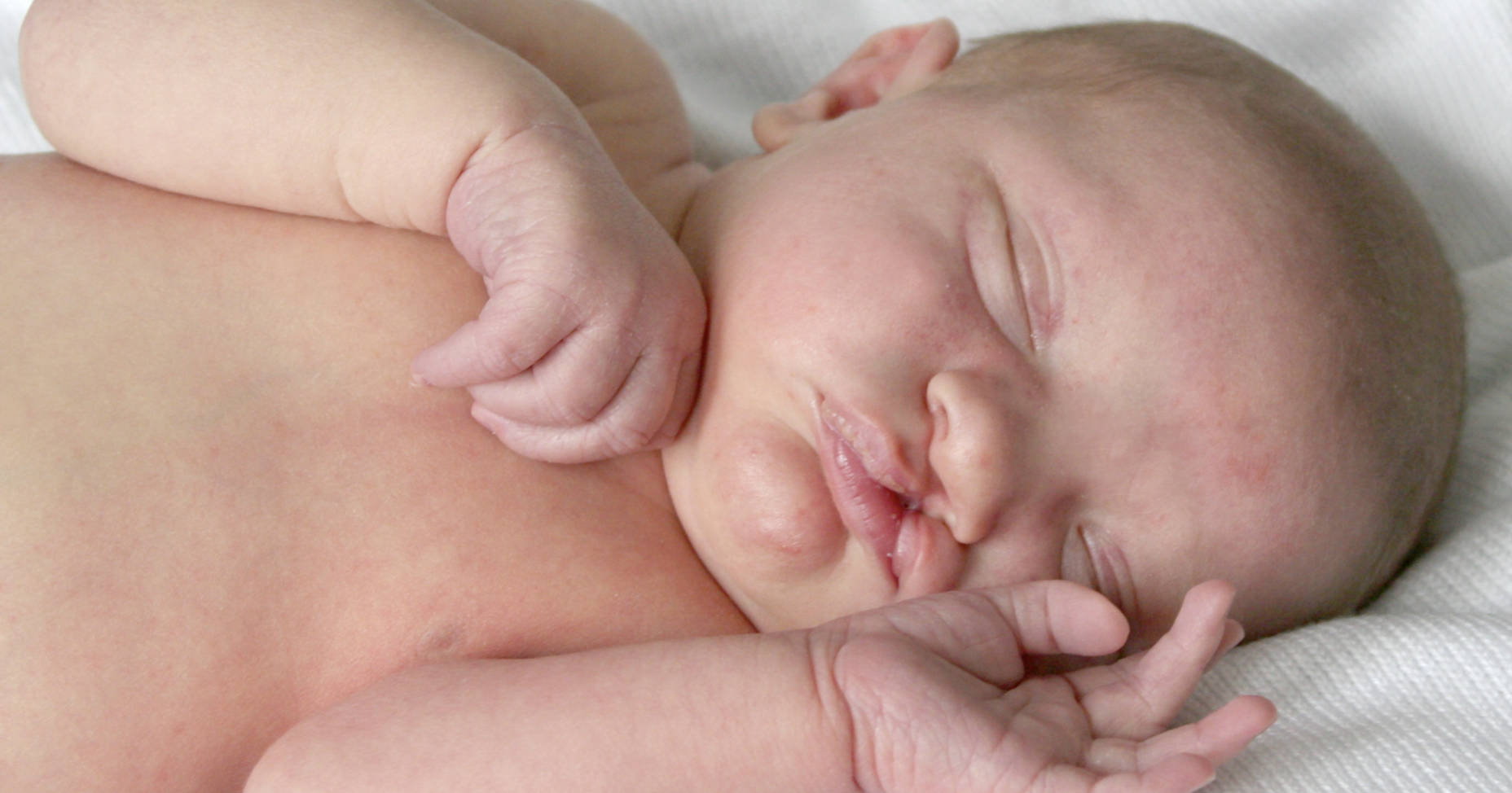
January is Birth Defects Awareness Month. Our skilled clinicians are dedicated to delivering expert care for women, babies and children — including preventing, diagnosing and treating an array of abnormalities present at birth or before.
Birth defects are structural changes present at birth that can affect almost any part of the body. Advancements in medicine and surgery have led to better survival and, thankfully, more children born with birth defects grow up to lead full lives.
We sat down with Amber Wood, M.D., a maternal-fetal medicine (MFM) specialist with Maternal-Fetal Medicine Specialists of Puget Sound, part of Pediatrix® Medical Group, and Paulette Abbas, M.D., a pediatric surgeon with Pediatrix Surgical Associates in Texas, to learn more about how birth defects are diagnosed and treated.
What are some of the most common birth defects you see in your practice?
Dr. Wood: As an MFM doctor, birth defects are one of the most common reasons we may see a patient for evaluation. Some of the most common birth defects we see in our practice include birth defects of the spinal cord (such as a neural tube defect), kidney anomalies, limb anomalies (such as clubfoot), cleft lip or palate, abdominal wall or bowel defects or cardiac defects. We also see and care for patients with rare birth defects.
Dr. Abbas: The most common birth defects we see that require surgery include congenital diaphragmatic hernia, gastroschisis, omphalocele, esophageal atresia, anorectal malformations/imperforate anus and Hirschsprung’s disease.
How are these defects diagnosed? What tests do you perform?
Dr. Wood: During pregnancy, birth defects are typically diagnosed by ultrasound. Some more severe birth defects may be able to be diagnosed during the 12- or 13-week ultrasound. However, most will be diagnosed during the detailed anatomy scan, typically performed at 20 weeks. If a birth defect is suspected on an ultrasound at an obstetrician-gynecologist’s (OBGYN) office, it’s important to have that ultrasound repeated at the MFM office since our sonographers and doctors are specially trained to diagnose and evaluate birth defects via ultrasound. If a birth defect is confirmed, the MFM specialist will speak with the OBGYN to review the findings and discuss the next steps in care.
Depending on the ultrasound findings, further studies may be recommended, such as a detailed ultrasound of the fetal heart (a fetal ECHO) or a fetal MRI (especially if there is concern about a spinal or brain defect) to help us gain more information. Other birth defects may be monitored on ultrasound throughout the pregnancy.
Since some birth defects may be linked to genetic causes, we suggest parents have a counseling session with a genetic counselor. A genetic screening or other testing may be conducted. Genetic screenings may include a blood draw, where testing can include a chorionic villus sampling (taking a sample of the placenta to evaluate fetal genetics, performed at 12 to 13 weeks gestation) or an amniocentesis (taking a sample of the amniotic fluid to evaluate fetal genetics, performed 16 weeks or greater). The genetic counselor reviews these options with parents and discusses what information might be learned from genetic screening or testing.
Q: How are common birth defects treated?
Dr. Wood: Some birth defects may never require treatment. If treatment is required, most birth defects are treated after the baby is born. For some major birth defects, surgery may be required soon after delivery. For other birth defects, immediate treatment may not be needed, and the baby can be monitored as he or she grows. All options are reviewed with parents at the time of diagnosis.
We monitor ultrasound findings throughout the pregnancy. We also arrange a consultation with the pediatric providers who will care for the baby after delivery. This helps parents know what to expect for postnatal care. For example, suppose there is a cardiac defect that would require treatment after delivery. In that case, the patient will meet with the pediatric cardiologist before delivery to review ultrasound findings and discuss plans for after delivery. Likewise, if a baby has an abdominal wall defect requiring surgery after delivery, parents meet with pediatric surgeons before delivery to review what that surgery will entail. The type of care needed after delivery depends on the organ system involved and the severity of the defect.
There are rare birth defects that, in certain cases, can be treated during pregnancy with fetal surgery (such as a neural tube defect). These cases are only performed at specialized centers and require that specific criteria are met for both the patient and the baby. If a patient and their baby are candidates, we help arrange an evaluation at one of the specialized centers.
Q: How can women reduce their baby’s risk of birth defects?
Dr. Wood: It’s important to know that there is a baseline risk of a major birth defect in 2% to 4% of all pregnancies, so they may occur even if you don’t have any known risk factors. Birth defects may also be secondary to genetic abnormalities or environmental exposures, although the exact cause is often unknown.
However, there are some steps patients can take to reduce their risk as much as possible, including:
- Get enough folic acid, even before conception. All women of reproductive age should take 400mcg of folic acid daily, even before trying to become pregnant, since the fetal spine and brain form at three to four weeks of gestation (typically before most people even know that they are pregnant). Adequate folic acid supplementation can help decrease the risk of birth defects of the spine and brain. You can get folic acid by taking a vitamin, eating fortified foods or a combination of both.
- Avoid alcohol while attempting to conceive and while pregnant. There is no known safe amount of alcohol during pregnancy, and the Centers for Disease Control and Prevention (CDC) recommends abstinence while attempting to conceive and during pregnancy. Drinking alcohol during pregnancy may be associated with abnormal facial features and neurodevelopmental disorders.
- Control diabetes before getting pregnant to minimize the risk of your baby developing birth defects. Women with a higher HbA1c level have a higher risk of birth defects (most commonly of the spine or heart).
- Discuss any medications you’re on with your doctor before getting pregnant. Women with chronic medical conditions that require long-term medication, such as lupus, hypertension, epilepsy, organ transplantation and diabetes, should have a preconception visit with the provider who manages their medications, as well as a visit with an MFM physician. While most medications are safe to use before and during pregnancy, some are associated with birth defects. Having a preconception visit allows for a medication review and, if needed, patients can be transitioned to a safer medication.
Q: Is there anything else you think is important for parents to know about birth defects?
Dr. Wood: Ultrasound is our main screening tool for diagnosing birth defects. The most important ultrasound is the 20-week anatomy scan. However, I also recommend an early anatomy scan at 12 to 13 weeks since some severe birth defects can be diagnosed at this time. Early diagnosis of a birth defect allows us to prepare for care after birth, gives parents an idea of what to expect for care after delivery of the baby and enables us to make sure that the delivery will take place in a location where the appropriate pediatrics teams are available to care for and support the baby after delivery.
It’s also important to know that there are limitations to our ability to diagnose birth defects before delivery. Minor birth defects may not be visible on ultrasound and may not be noted until after birth. In addition, in some cases, it can be difficult for us to know how a birth defect may affect a baby after delivery. For example, a mild to moderate birth defect in the brain may have a normal outcome or cause a developmental delay. We are often unable to say for certain in some cases until after the baby is born and has a neurologic exam.
Q: What advice do you have for parents?
Dr. Wood: If you or your partner have a known family history of birth defects or genetic disorders, please let your OB providers know. A consultation with a genetic counselor can be arranged, and we may offer earlier ultrasounds or specific genetic testing if desired.
We understand that hearing that there is a concern for a birth defect is almost always unexpected and difficult news to learn. We do our best to provide compassionate, supportive and informed care for our patients in these unexpected situations.
It is also important to know that in most cases, birth defects happen sporadically and are not related to something that the parents could have prevented. Hearing unexpected news about a birth defect is always understandably hard for parents, and it is important to know that it is not your fault this happened!
Dr. Abbas: This is still your precious baby, and none of these diagnoses or surgeries change that. It may not be how you envision your postnatal period but try to enjoy the arrival of your sweet child. Here is my advice:
- It’s important to lose the mom guilt! As a mother, I know it’s easy for us to blame ourselves for anything that happened. However, these anomalies did not occur because of anything you did, ate, or wore during pregnancy.
- Ask questions. This can be overwhelming, and often so much information is given at once, which can be hard to process. Don’t be embarrassed to ask the same questions again, and write down your questions (and the answers).
- It always feels overwhelming at the beginning, but every parent can adapt and adjust. You will be able to do what it takes for your baby, no matter how unimaginable that seems in the beginning.
- Be sure to take care of yourself during this time. It can be easy to forget to eat or sleep when your child is in the hospital, but you must remember to stay healthy for both yourself and your baby.
Remember that your whole medical team has the same goal — to get your precious child home as quickly and safely as possible. So please use us as a resource.
FAQs About Common GI Defects
Q: What is a congenital diaphragmatic hernia (CDH), and how is it treated?
Dr. Abbas: CDH is a hole in the muscle of the diaphragm, the structure that separates your chest from your belly. It is involved in breathing. This hole develops during pregnancy and allows abdominal contents (intestines, stomach, spleen, liver) to go up into the chest. This causes the lungs to be squeezed during pregnancy and limits the development and growth of the lungs. The hernia can also cause tightness in the vessels going to the lungs, which can affect blood flow to the lungs.
When a baby is born with CDH, the most important aspect of care is ensuring the lungs are working well and stable. Often, this requires a breathing machine and a few different types of medications and, as a last resort, may even include a heart-lung bypass machine called ECMO. Once the baby is stable and the lungs are strong enough, the baby can undergo surgery to fix the hernia. This can be done a variety of ways — open incision or with a camera through the chest or abdomen — but the basic goals are the same: to pull down the abdominal contents and close the hole in the diaphragm. Generally, no further surgery is required for the hernia after repair, but there is a chance of the hole opening back up.
Q: What are abdominal wall defects, and how are they treated?
Dr. Abbas: Gastroschisis and omphalocele are abdominal wall defects. Gastroschisis is when the intestines poke through a defect next to the belly button and is directly visible. Omphalocele is when the intestines poke through a defect in the belly button and is often covered with a membrane. The treatment for gastroschisis is to reduce the bowel back into the abdomen and use the umbilical cord as a flap to cover the hole. However, if the bowel is unable to safely go back into the abdomen, a silo is placed. This is a special plastic bag that is placed on the abdomen to contain the intestine poking out. Over the next few days, the bag is gently squeezed from the top down to guide the intestines into the abdomen slowly. Once the intestines are back in, the silo is removed, and the hole is closed. Omphaloceles are usually managed by leaving the membrane intact and “painting” it to cause it to thicken and develop a thick, skin-like covering. The resulting hernia can be fixed in the future. If the omphalocele is small, the membrane can be removed and the defect closed.
Q: What is esophageal atresia, and how is it treated?
Dr. Abbas: Esophageal atresia occurs when the esophagus (the feeding tube connecting the mouth to the stomach) doesn’t connect, so there are upper and lower sections that don’t connect. An abnormal connection between one or both pouches to the trachea (the windpipe) can damage the lung if intestinal contents enter the trachea and go into the lungs.
Surgery is required soon after the patient is born to disconnect any abnormal connections and reconnect the esophagus. However, sometimes, this is not possible if the patient is sick or if the ends of the esophagus are too far apart. If that is the situation, the patient may have a gastrostomy tube (a tube in the stomach) surgically placed to drain the stomach contents away from the lungs.
Once the child is more stable or bigger, corrective surgery can be performed. This involves disconnecting the abnormal connection, closing the trachea hole is closed, and sewing the esophagus ends together.
Q: What is intestinal atresia, and how is it treated?
Dr. Abbas: During a baby’s development, parts of the bowel can develop an internal scar or wall that causes a blockage, and some don’t connect. This blockage, called intestinal atresia, prevents the contents of the intestines from appropriately moving down and can cause an obstruction where the contents of the intestines can’t adequately pass through. When this happens, the contents flow back up and the patient becomes bloated, vomits, and has decreased stools. These atresias can occur in any part of the intestines.
Surgery is used to remove the blockage and reconnect the intestines. The method of reconnection depends on the location. After surgery, the area where the bowel was reconnected can scar and cause another obstruction. In this case, another surgery may be required.
Q: What is an anorectal malformation/imperforate anus, and how is it repaired?
Dr. Abbas: Sometimes babies are born without an anus to poop through. Sometimes there may be a very small hole near where the anus might be (called a fistula), but there is no well-defined anus to allow the baby to poop normally.
The patient often requires immediate surgery with a colostomy if there is no fistula/small hole. If a fistula is present, it can often be stretched to allow the patient to poop temporarily. Ultimately, babies with this condition often require surgery to create an anus in the normal location.
Q: What is Hirschsprung’s disease, and how is it treated?
Dr. Abbas: The intestine has a series of nerves that send signals to the bowel to relax and allow for normal movement. These nerves develop starting at the beginning of the intestines and continue developing down to the rectum. However, in some patients, the nerves stop developing at a certain point, and all the bowel past that section has no nerves. This causes the section of the bowel without nerves to be in a constant state of contractions, making bowel movements difficult.
Rectal dilations and irrigations are the first steps to treat Hirschsprung’s disease. They allow the contracted bowel to release its contents and prevent bacterial overgrowth and infection. Then, the patient will undergo a biopsy to find where the nerves stopped growing (called the level). Once the level is identified, there are two surgical options. The type of surgery depends on the patient’s age, the length of the bowel involved and the presence of any other medical issues.
